Colonoscopy reveals mucosal edema, erythema, granularity, and decreased vascular pattern in the entire colon, with relative rectal sparing. The terminal ileum appears normal. Some representative images are shown.
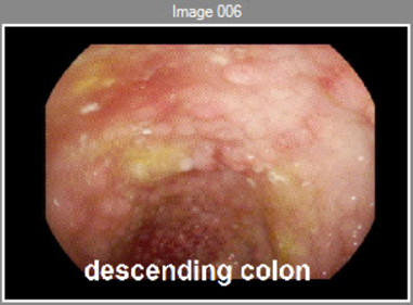
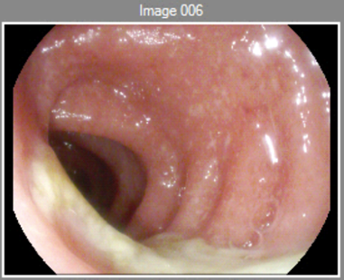
Pathology shows chronic active colitis. There are no granulomas, viral cytopathic changes, or dysplasia.
What additional evaluation would you order after performing the colonoscopy? Click to see the results.
CBC
Hgb 11.5, low-normal MCV
CMP
Normal Cr and LFTs
Serum inflammatory marker(s)
ESR 30, CRP 11
Stool infectious studies
C. difficile negative, stool culture negative, ova and parasite negative
TPMT activity
TPMT activity normal
TB test
Quantiferon negative
Hepatitis B serologies
Hepatitis B surface Ab positive, surface Ag negative, core Ab negative
MR enterography
MR enterography shows wall thickening and mucosal hyperemia of the entire colon except for the rectum, and no evidence of small bowel disease.
Click here to move on to the final discussion
Click here to return to the previous part