Case 14.5
The patient was referred to an endoscopist with expertise in chromoendoscopy.
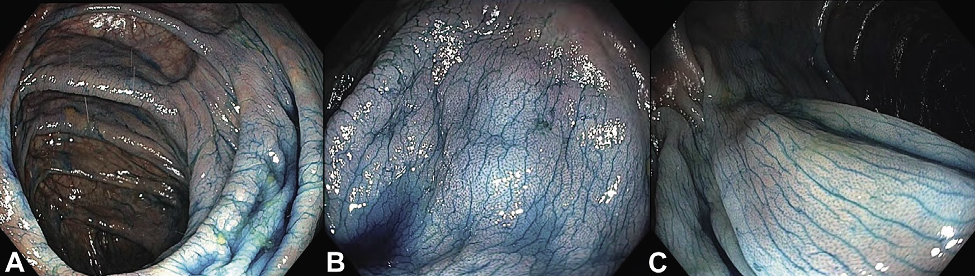
Chromoendoscopy uses a dye solution of either indigo carmine or methylene blue onto the colonic mucosa to provide contrast enhancement to augment visualization of epithelial surface detail during colonoscopy. The innominate grooves of the normal colon in a patient with ulcerative colitis can be easily seen with the use of dye, facilitating efficient examination of the surface during colonoscopy surveillance for dysplasia.
The endoscopist examined the colon using diluted indigo carmine pan chromoendoscopy and found a 16 mm sessile serrated lesion in the transverse colon. The endoscopist removed the lesion by hybrid endoscopic submucosal dissection en bloc as seen in the figure below.
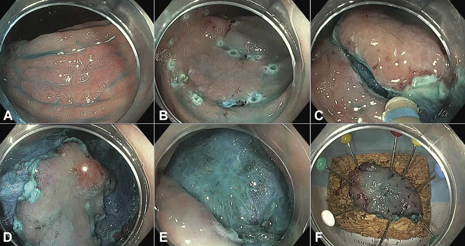
A. The lesion and its borders are assessed. B, The periphery of the lesion is marked, and the lesion is injected using dynamic submucosal injection. C, Circumferential incision. D, Some submucosal dissection. E, The lesion is ultimately resected en bloc using a stiff snare. F, The specimen is pinned for orientation and histologic assessment.