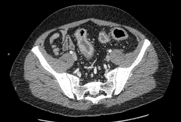
CT A/P with contrast:
Wall thickening and mucosal hyperemia of the rectosigmoid colon, most consistent with active inflammation/colitis. No abscess, no bowel obstruction, and no significant inflammatory changes of the terminal ileum.
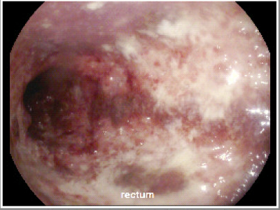
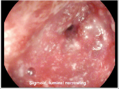
Colonoscopy:
Severe inflammation throughout the rectum and visualized sigmoid colon, characterized by severe erythema, spontaneous bleeding and friability, and diffuse ulcerations. There was an area of luminal narrowing at 30 cm from the anal verge that was not passable with the adult colonoscope, so the procedure was aborted after biopsies were taken.
Click here to move on to the next part
Click here to return to the previous part