Computed tomography (CT) of the abdomen and pelvis was ordered:
Mild wall thickening and mucosal hyperenhancement of the rectum, sigmoid, descending, and transverse colon. The ascending colon and cecum are normal. The small intestine is normal.
Colonoscopy was performed and revealed:
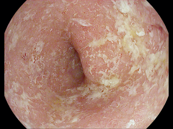
Diffuse edematous, erythematous mucosa with friability and erosions within the rectum, sigmoid colon, and descending colon up to 60cm (image below.), the remainder of the colon was normal. Cold forceps biopsies demonstrated chronic active colitis without granulomas or dysplasia.

How would you grade the above image?
Mayo 2 Colitis
The erythema and loss of vascular pattern in this image are consistent with Mayo 2 colitis
Mayo 1 colitis
Mayo 3 colitis
Click here to move on to the next part
Click here to return to the previous part
